19. Week 3 of Development: The Notochord, Neural Tube, and Allantois
Review of MEDICAL EMBRYOLOGY Book by BEN PANSKY, Ph.D, M.D.
- Notochord development: in the human, the notochord is a cellular rod that develops from the prochordal process and forms the first longitudinal midline axis around which the vertebral bodies are organized and is the basis for the axial skeleton. It will later regress. By day 12 or 13, the notochord is visible throughout the length of the embryo and around it are layered concentrations of cells, representing the primordia of the future vertebral bodies
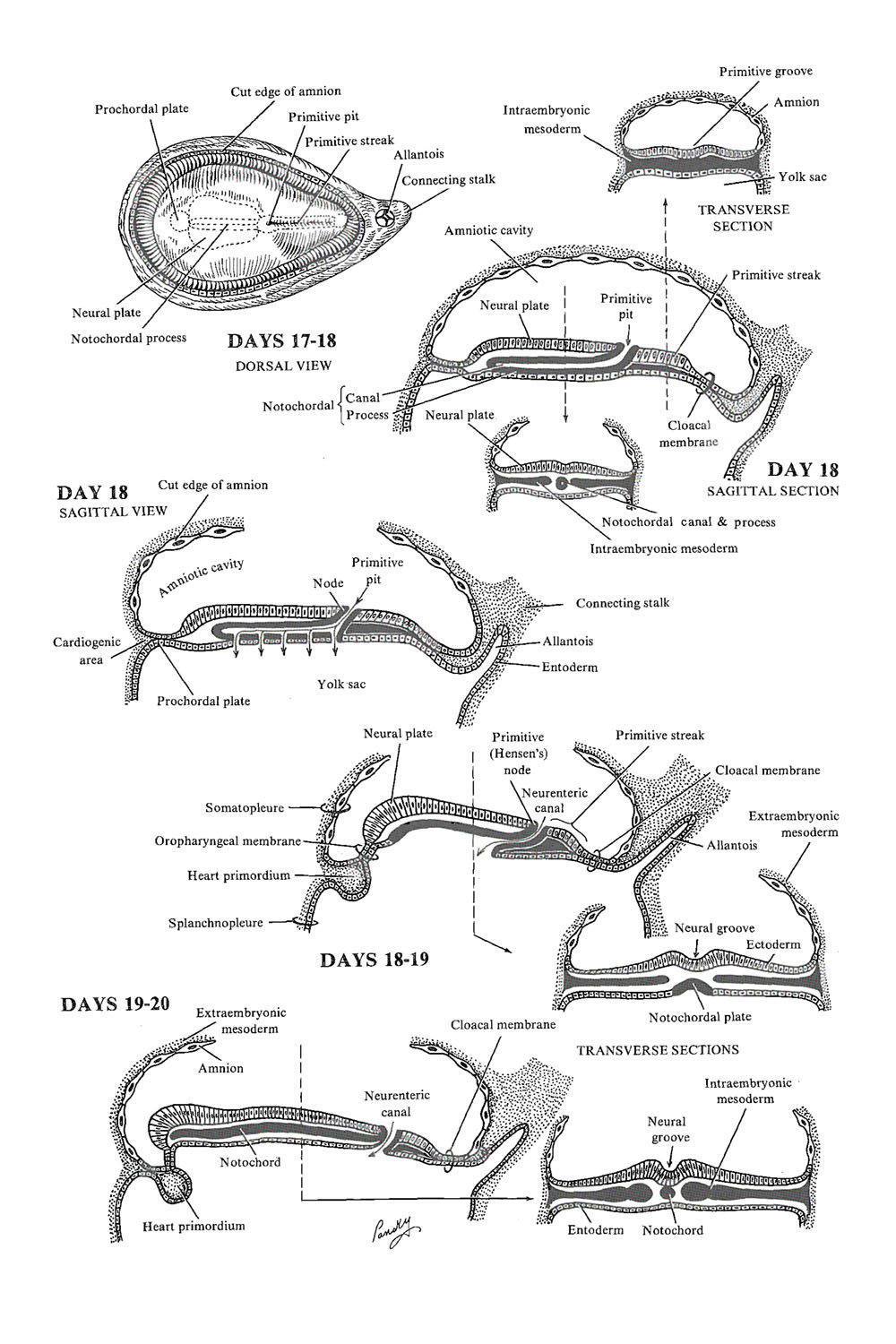
- STAGE OF THE NOTOCHORDAL PROCESS (entire area cephalic to the primitive streak): about day 17
- The floor of the notochordal process fuses with the underlying endoderm as it undergoes preferential growth. Hensen's node seems to recede toward the caudal end
- PROCHORDAL STAGE: about day 19
- Degeneration of the fused region takes place, and openings appear in the floor of the notochordal process (resorption of the floor), opening a communication between the yolk sac and the notochordal canal, a lumen which is formed as the primitive pit extends into the notochordal process during its development
- The openings become confluent, and the floor of the notochordal canal disappears. A small passage, the neurenteric canal, temporarily connects the yolk sac and the amniotic cavity
- NOTOCHORD STAGE: about day 20
- The notochord process remains and forms a grooved, flattened plate, the notochordal plate, which, beginning at its cranial end, infolds to form the notochord. The embryonic endoderm again forms a continuous layer below the notochord The latter is thus the primary skeleton of the 3-layer embryo
- Neural tube development (neurulation)
- THE EMBRYONIC ECTODERM over the developing notochord thickens to form a neural plate (about day 18) which apparently is enduced by the developing notochord and paraxial mesoderm on either side
- The plate first appears cranial to the primitive knot and dorsal to the notochordal process with mesoderm adjacent to it
- With the elongation of the notochordal process, the neural plate broadens and extends cranially to the oropharyngeal membrane
- The ectoderm of the plate is called neuroectoderm and eventually gives rise to the central nervous system (brain and spinal cord)
- THE NEURAL PLATE, on about day 20, invaginates along its central axis to form the neural groove with neural folds created on each side of the groove
- By the end of week 3, the neural folds move together, fuse, and convert the neural plate into the neural tube. Closure begins in the middle of the embryo and progresses toward both cephalic and caudal ends. It begins on day 21
- Closure of the neural groove is more rapid toward the cephalic (anterior) end than toward the caudal (posterior) end
- The anterior or cranial neuropore closes in week 4 (day 26), whereas the posterior or caudal neuropore closes near day 28
- Neuroectodermal cells at the lateral edge of the neural plate -do not become part of the tube but form a neural crest over the neural tube and give rise to the neural crest cells
- The allantois: appears on day 16 as a small, fingerlike outpouching or diverticulum from the caudal wall of the yolk sac. It remains small in the human embryo, is involved with early blood formation, and is related to the development of the urinary bladder