47. Development of The Mesenteries
Review of MEDICAL EMBRYOLOGY Book by BEN PANSKY, Ph.D, M.D.
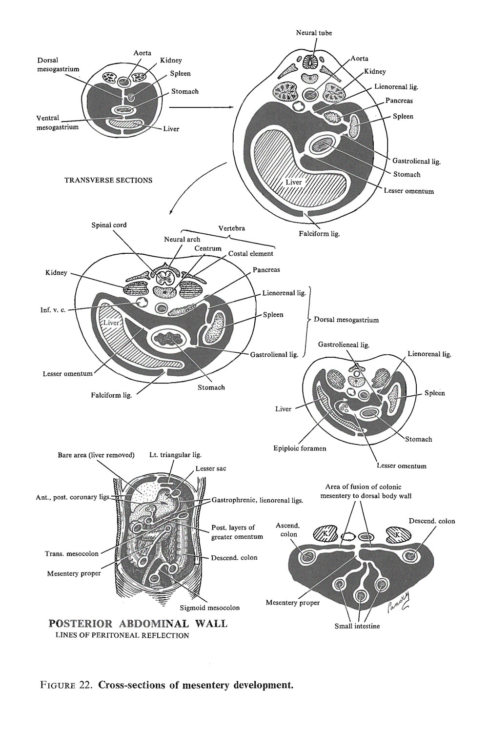
- Introduction: the 2 apposing splanchnic mesoderm layers fuse to form a double-layer membrane, the primitive mesentery, with the closing of the abdominal wall and the formation of the peritoneal cavity
- THE VENTRAL MESENTERY
- With formation and closure of the abdominal wall, the stomach and upper duodenum, which are initially in contact with the septum transversum, draw away from the septum, except for their ventral borders which stay connected to the underside of the septum and the anterior abdominal wall by the ventral mesentery
- During formation of the ventral mesentery, the liver grows so rapidly that the septum transversum can no longer accommodate it, so it protrudes between the 2 leaves of the mesentery, dividing the latter into an anterior part, the falciform ligament (from liver to anterior abdominal wall) and a posterior part, the lesser omentum (between the liver and ventral borders of the stomach and duodenum)
- The free edge of the falciform ligament contains the umbilical vein which is obliterated after birth to from the ligamentum teres hepatis (round ligament)
- The free edge of the lesser omentum contains the common bile duct, the portal vein, and the hepatic artery
- Initially, the lesser omentum is oriented in a sagittal plane, but with rotation of the stomach and growth of the liver, it acquires a frontal position so that its lower edge forms the upper margin of the epiploic foramen of Winslow - the entrance into the lesser peritoneal sac behind the stomach
- The liver is completely surrounded by ventral mesentery except on its upper surface where it adjoins the diaphragm ? the so-called bare area of the liver
- The lines of reflection, where peritoneum over the liver is continuous with that on the underside of the diaphragm, are called the coronary ligaments
- THE DORSAL MESENTERY extends from the lower esophagus to rectum and throughout its length serves as a pathway to the gut for blood vessels, nerves, and lymphatics
- In the area of the duodenum, it is called the dorsal mesoduodenum
- Rotation of the stomach and duodenum with growth of the head of the pancreas, causes the duodenum to swing from midline to the right side of the peritoneal cavity, pushing the duodenum and pancreatic head against the dorsal body wall and fusing the right surface of the dorsal mesoduodenum with wall peritoneum
- Both layers disappear completely, except in the area of the pyloris of the stomach, where a small part of duodenum remains intraperitoneal, and the duodenum and pancreatic head become fixed in a retroperitoneal position
- In the area of the colon, it is called the dorsal mesocolon
- When the ascending and descending colons acquire their definitive positions, their mesenteries are pressed against the abdominal wall peritoneum, and the colons are permanently anchored or fused as retroperitoneal. The appendix and lower end of the cecum, however, retain a free mesentery
- The transverse mesocolon, at first, covers the duodenum with an additional peritoneal layer, but later fuses with the posterior wall of the omental burs Its final lines of attachment extend from the hepatic to the splenic flexures
- In the area of the stomach, it is called the dorsal mesogastrium or greater omentum
- The dorsal mesentery of the jejunum and ileum is the mesentery proper which changes with rotation and coiling of the intestinal loops
- When the lower limb of the primitive intestinal loop moves to the right side of the abdominal cavity, the dorsal mesentery twists around the origin of the superior mesenteric artery
- The mesentery of the jejunoileal loops is initially continuous with that of the ascending colon, but when the ascending mesocolon fuses with the posterior abdominal wall, the mesentery of the loops obtains a new line of attachment, extending from where the duodenum is intraperitoneal to the ileocecal junction