126. Development of The Venous System: The Inferior Vena Cava
Review of MEDICAL EMBRYOLOGY Book by BEN PANSKY, Ph.D, M.D.
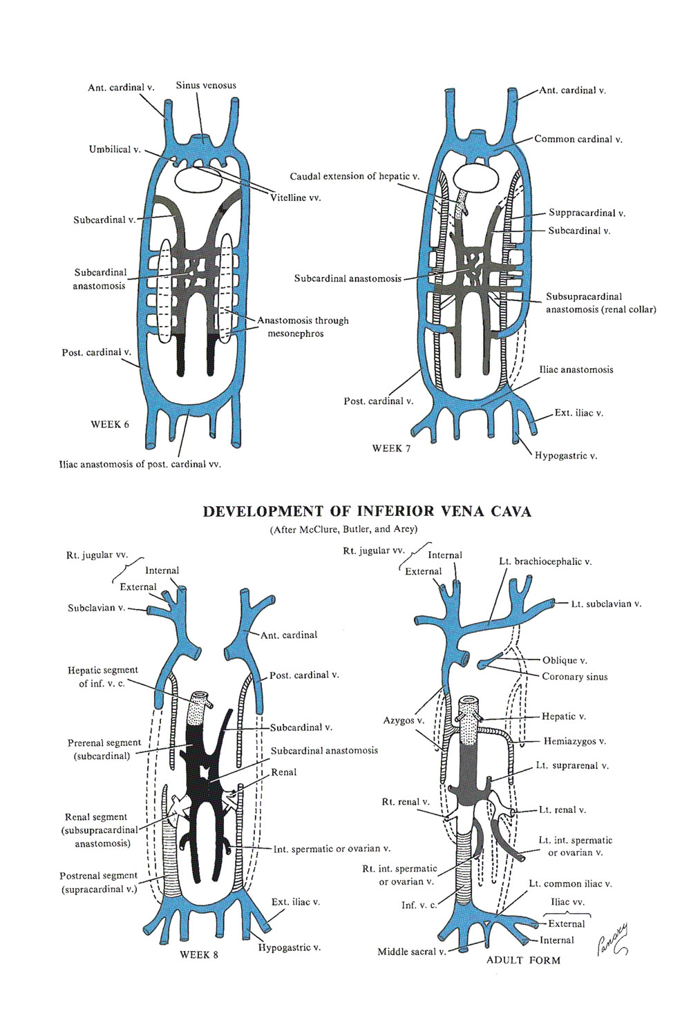
- The inferior vena cava: a series of successive venous networks take part in the formation of the inferior vena cava. Each predominates temporarily, then regresses, and remains only partly in the final definitive system
- THE MESONEPHROS grows considerably and becomes very highly vascularized during week 4. Although it is drained initially only by the posterior cardinal veins, a new system takes over after week 4, the subcardinal network, which is formed by the internal veins of the wolffian body
- The internal veins of the wolffian body are widely anastomosed with the initial posterior cardinal network and with each other, forming the median subcardinal network, which soon predominates. It takes over the posterior cardinal system, which disappears in the middle region of the embryo
- The subcardinal sinus persists as the left renal vein
- The anterior segment of the left subcardinal vein disappears, but its posterior segment forms the left gonadal vein
- The right subcardinal vein forms the right gonadal vein and the pararenal portion of the definitive inferior vena cava
- Cranially, it continues with the mesenteric segment and the hepatic segment derived from the hepatic vein (proximal right vitelline} and hepatic sinusoids
- DURING WEEKS 6 AND 7, a supplementary dorsal network develops, called the supracardinal system, which runs parallel to the paravertebral sympathetic chain and opens into the proximal segment of the posterior cardinal veins. Anastomoses are formed
- Between the two supracardinal veins
- Between the supracardinals and the subcardinals, on the right side
- Between the extremities of the posterior cardinal veins
- The left supracardinal vein becomes the hemiazygos vein and is drained toward the right by the transverse anastomosis which forms an interazygos communication
- The right supracardinal vein becomes the azygos vein, which opens into the right anterior cardinal vein
- From below, the azygos vein drains the 2 iliac veins, thus becomes the prerenal portion of the definitive inferior vena cav
- IN SUMMARY: the definitive inferior vena cava is composed of (from caudal to cranial)
- The posterior intercardinal anastomosis
- The caudal portion of the right supracardinal vein
- The right anastomosis between the supracardinal and the subcardinal veins
- A segment of the right subcardinal vein
- The anastomosis between the right subcardinal and right vitelline veins
- The terminal portion of the right vitelline vein
- Malformations of the inferior vena cava
- MALFORMATIONS of the inferior vena cava may be due to the complexity of formation of the system, yet, even in cases of severe malformation, one of the constituent networks invariably substitutes some form of venous flow
- Agenesis (absence) of the inferior vena cava is the most conspicuous. The right subcardinal vein has failed to make its connection with the liver and shunts blood directly into the right supracardinal vein. Thus, blood from the caudal part of the body reaches the heart via the azygos and superior vena cav The hepatic vein enters the right atrium at the site of the inferior vena cava
- An abnormality of position of the vein may affect the adjacent organs, such as the ureter, compressing it and causing a hydronephrosis
- Double inferior vena cava at the lumbar region: the left sacrocardinal vein has failed to lose its connection with the left subcardinal, and the left common iliac vein may or may not be present. The left gonadal vein is usually present and in a normal condition