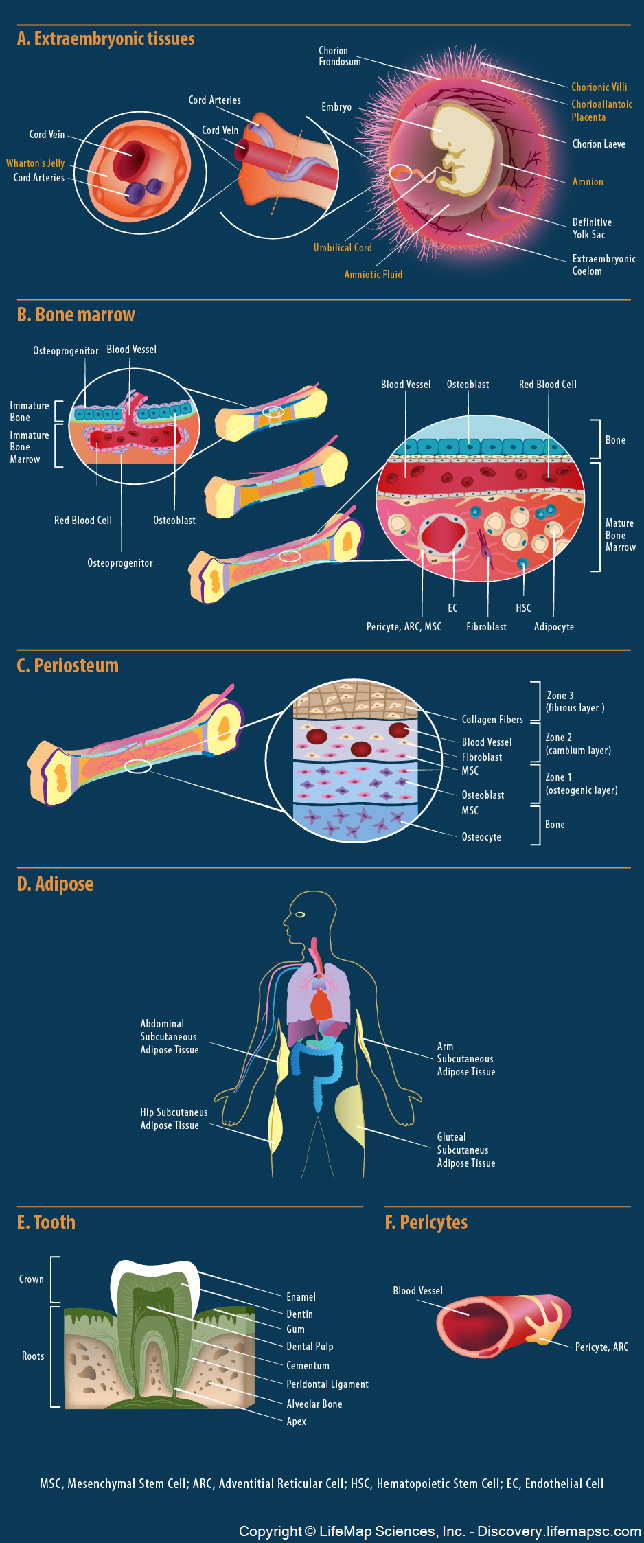
Schematic presentation of the main embryonic, fetal and adult origins of mesenchymal stem cells (MSCs):
- Extraembryonic tissues - during fetal development, MSCs can be found in multiple fetal tissue sources: the embryonic parts of placenta (i.e., chorionic villi), the amnion, which envelops the embryo and its fluid (i.e., amniotic fluid), and the umbilical cord, which contains the surrounding Wharton's jelly tissue and the cord vessels (two arteries and one vein).
- Bone Marrow - in the immature bone, osteoprogenitor cells in the periosteum (left panel; blue-purple cell) invade the forming bone marrow with the associated blood vessels to form the putative mature bone marrow stromal cell (right panel; i.e., pericyte, ARC, MSC).
- Periosteum - In the mature bone, the periosteum is a specialized connective tissue that forms a fibrovascular membrane covering bone surfaces (with the exception of the joints of long bones). The periosteum is divided into several layers or zones (starting from the outermost bone layer): the osteogenic layer, which contains osteoblasts and MSCs, the cambium layer which contains MSCs, fibroblasts and blood vessels and the fibrous layer, which contains collagen fibers and fibroblasts.
- Adipose - the white subcutaneous adult adipose tissue, found in several anatomical locations such as the arms, abdomen, hips and the gluteal region, is a source of adipose MSCs. Other human adipose depots, not depicted in the image, also exist and may contain similar MSCs.
- Tooth - from the early stages of tooth development ('deciduous teeth') to adult stages ('permanent teeth'), the innermost part of the tooth, termed the dental pulp, contains dental pulp stem cells (i.e., MSCs).
- Pericytes – Pericytes sit on the walls of blood vessels incorporated in various tissues (e.g., bone marrow and adipose). Specialized pericytes (on small blood vessels) and adventitial articular cells (ARCs; on large blood vessels) are assumed to be a major source of MSCs throughout human organs.


